

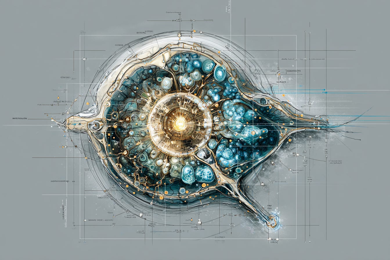
A pathologist examining a breast cancer biopsy can read a great deal from the slide. The cells — stained pink and purple — arrange themselves in patterns that decades of training make legible. Some areas are dense with malignant cells: the tumour’s centre. Others show the advancing edge, where cancer is pushing into healthy tissue. Some regions are infiltrated by immune cells; others appear almost entirely free of them.
What the traditional biopsy slide cannot show is what those cells are actually doing — which genes each one is expressing, what proteins it is making, what signals it is sending to its neighbours, why the immune cells in one region are actively attacking the tumour while the immune cells in another region appear to have gone quiet. The stain shows structure. It cannot show function.
For most of the history of molecular biology, researchers addressed this limitation by doing what they could: taking the tissue, grinding it up, and measuring the average gene expression of the resulting mixture. This approach called bulk sequencing produces useful information. It also, inevitably, hides the detail that matters. A tumour cell that is resistant to a drug and a tumour cell that is sensitive to the same drug look identical in a bulk sample. Their individual signals cancel each other out. What the measurement captures is the average, and in a heterogeneous tissue, the average misleads.
Single-cell and spatial biology exist to fix that problem. Single-cell technologies analyse gene expression at the resolution of individual cells. Spatial technologies add location: they preserve the tissue architecture and record not just what each cell is doing but where in the tissue it sits. Together, they produce something that did not exist before — a map of a living tissue that shows, at microscopic resolution, which genes are active in which cells in which locations, and what those cells are saying to each other.
Nature Methods named spatially resolved transcriptomics its Method of the Year in 2020. Since then, the technology has moved from the leading edge of basic research to commercial platforms available in well-equipped laboratories worldwide. In February 2025, Illumina announced a spatial technology product planned for commercial release in 2026 with a capture area nine times larger than existing technologies and four times greater resolution. The field has tipped from pioneering to mainstream. What that means for understanding disease — and ultimately for treating it — is the subject of this article.
The tumour is not the problem. The neighbourhood is.
The central insight that spatial biology has delivered to cancer research can be stated simply: a tumour is not a mass of identical malignant cells. It is an organised community of different cell types — cancer cells in various states, immune cells of multiple kinds, structural cells called fibroblasts, blood vessel cells, and others — all occupying distinct spatial niches within the tissue, interacting constantly with their immediate neighbours, and behaving very differently depending on where they are.
This was suspected for a long time. What spatial biology has done is make it visible and measurable.
One of the most striking findings from spatial transcriptomics research is that the same tumour can simultaneously contain cells that are highly sensitive to a treatment and cells that are highly resistant — and that this is not purely a matter of genetics. Location matters. A cancer cell sitting in the centre of a tumour, surrounded by other cancer cells and relatively few immune cells, behaves differently from a genetically identical cancer cell at the tumour’s leading edge, where it is in constant contact with immune signals and structural proteins from the surrounding healthy tissue. The edge cell may be more aggressive and invasive; the centre cell may be more metabolically active but less mobile. Their gene expression profiles differ. Their drug sensitivities differ. The treatment that kills one may leave the other intact.
A tumour contains cells that are sensitive to a drug and cells resistant to the same drug — not just because of genetics, but because of location. Cells at the leading edge behave differently from cells at the centre. Spatial biology makes this visible.
A 2023 paper in Nature Communications on oral squamous cell carcinoma demonstrated that the gene expression profile of cells at the tumour’s leading edge — what researchers call the leading edge signature — was consistently associated with worse clinical outcomes across multiple cancer types, while the tumour core signature was associated with improved prognosis. The spatial position of the cancer cells relative to the boundary between tumour and healthy tissue carries predictive information that no amount of bulk sequencing could have extracted, because bulk sequencing cannot preserve the information about where each cell was sitting.
Immune cells show equally striking spatial organisation. Immunotherapy — the class of treatments that effectively unlocks the immune system to fight cancer — works by removing the signals that cancer cells use to suppress immune activity. It works well for some patients and not at all for others. Spatial biology research is revealing why. In patients who respond to immunotherapy, spatial analysis shows specific arrangements of immune cells clustered in particular locations around the tumour — what researchers call immune hubs or tertiary lymphoid structures. In patients who do not respond, those structures are absent or differently arranged. The presence or absence of the structures is not detectable by any analysis that does not preserve the spatial information. A blood sample, a bulk biopsy, even conventional single-cell sequencing without location data cannot tell you whether the right immune cells are in the right places.
Spatial transcriptomics of small cell lung cancer tumours has identified two distinct subtypes of cancer cells within the same tumours: one associated with treatment resistance and high proliferative activity, the other associated with treatment sensitivity and greater immune cell contact. The resistant subtype appears to reshape its local environment to drive away immune cells — to domesticate the macrophages in its vicinity and convert them into accomplices in suppressing the immune response. This finding was invisible to conventional analysis. It required spatial mapping of the tumour to become apparent.
The platforms are ready. The clinical pipeline is not.
The commercial field for spatial biology has developed quickly and is now genuinely competitive. 10x Genomics offers the Xenium In Situ platform, which can detect up to 5,000 genes across tissue samples at subcellular resolution. Bruker’s CosMx platform can profile over 19,000 genes — the entire protein-encoding transcriptome. Visium HD, also from 10x Genomics, resolves spatial data to 2-micrometre bins. Illumina’s forthcoming platform will use a capture area nine times larger than current technologies, integrated with its Connected Multiomics software suite for analysis.
Key platforms:
spatial and single-cell biology (2026) — 10x Genomics Xenium In Situ — Up to 5,000 genes; subcellular resolution; widely adopted clinically
Bruker CosMx — Over 19,000 genes (whole transcriptome); single-cell resolution
10x Genomics Visium HD — 2µm spatial resolution; whole-transcriptome; sequencing-based
Illumina Spatial (2026 launch) — 9× larger capture area; 4× higher resolution; NovaSeq compatible
Vizgen MERSCOPE — MERFISH-based; high-plex; single-molecule resolution in intact tissue
STOmics Stereo-seq — 500nm resolution; large tissue sections; strong in developmental biology
Chan Zuckerberg Initiative / 10x Genomics / Ultima Genomics
Billion Cells Project: single-cell dataset of one billion cells launched February 2025 to train AI models for biology
The availability of these platforms has shifted the technology from something that required specialist physics laboratories to something that any well-resourced research institution can operate. The research output has followed. A review of spatial omics in cancer research published in Cancer Cell in January 2026, by researchers at the MD Anderson Cancer Center, described how spatial technologies now map tumour architecture with a fidelity that earlier methods could not achieve, resolving functional niches and spatial communities, converting spatial patterns into mechanistic insights.
The transition from research tool to clinical instrument, however, has several steps remaining. The data volumes generated by spatial transcriptomics are enormous. A single spatial experiment can produce hundreds of gigabytes of data. Analysing it requires computational infrastructure and bioinformatics expertise that most clinical settings do not yet have. The integration of spatial transcriptomics data with conventional pathology, with genomics, with clinical records, and with the kind of standardised workflows that regulatory agencies require before a diagnostic test can be approved — all of this is in progress but not yet complete.
The cost has been falling but remains significant. Running a spatial biology experiment on a clinical sample costs more than conventional sequencing and takes longer. For research settings, this is manageable. For routine clinical use — for the pathologist examining a cancer biopsy to guide treatment decisions for a patient who needs an answer within days — the workflow, cost and analytical requirements are still a barrier. The question of where spatial biology first enters clinical practice is the most practically important one in the field right now.
The most plausible near-term pathway is through what researchers call companion diagnostics: tests that are performed alongside a specific treatment to determine who is most likely to benefit. The patterns that spatial biology can detect — the presence or absence of immune structures around a tumour, the proportion of cells with certain gene expression signatures at the tumour edge, the spatial arrangement of drug-resistant subpopulations — are exactly the kind of information that could guide decisions about whether to use immunotherapy, which chemotherapy to start with, or whether a tumour is likely to respond to a newly approved targeted agent.
Why drug resistance looks different from here
Drug resistance is the central unsolved problem of cancer treatment. Most patients who are initially helped by a drug eventually stop responding to it. Understanding why, and predicting in advance who will stop responding and when, is the problem that determines whether a patient can be switched to an effective treatment before their cancer becomes untreatable.
Spatial biology is offering new ways to think about this problem. The conventional model of drug resistance focuses on genetics: a cell acquires a mutation that makes it insensitive to the drug, and that cell proliferates while the drug kills everything else. That model is real and important. But spatial biology research is demonstrating that it is incomplete. Resistance is also ecological.
A resistant cell does not only survive because of its own genetic properties. It also survives because of where it is in the tissue — who its neighbours are, what signals they are sending, whether the local immune environment is permissive or hostile. Research in melanoma has shown that distinct resistant fates — different ways a cell can become drug-resistant — are not randomly distributed through a tumour. They are spatially organised, associated with specific microenvironments. The cells in regions of necrosis behave differently from the cells adjacent to functional blood vessels. The resistant fate a cell adopts appears to be partly determined by its location.
This has immediate implications for treatment design. If resistance is partly spatial, then treating a tumour as a single uniform target with a drug calibrated to the average cell will predictably fail. The cells most resistant to treatment are often those in locations that the drug reaches least effectively and where the microenvironment most actively suppresses the immune system. A treatment strategy informed by spatial biology might target not just the cancer cells but their supporting environment: the macrophages they have domesticated, the fibroblasts that form barriers, the immune exclusion zones that protect the most resistant subpopulations.
If resistance is partly spatial, treating a tumour as a uniform target will predictably fail. The cells most resistant to treatment are often in the locations least accessible to drugs and most protected by their microenvironment.
Spatial analysis of breast tumours combined with pharmacogenomic profiling has identified that cancer cells in immunosuppressive microenvironments — the regions of the tumour where the immune system has been most effectively shut down — show specific sensitivity to cell cycle arrest agents and a class of drugs targeting the PI3K/AKT/mTOR pathway. This is information that could not be derived from bulk sequencing, because the immunosuppressive microenvironment and the cancer cells within it cannot be distinguished in a ground-up tissue sample. Seeing the map reveals what the average conceals.
Beyond cancer, spatial biology is beginning to reveal the tissue architecture of conditions that have been poorly understood precisely because they are diseases of organisation rather than diseases of individual cells. Pulmonary fibrosis — scarring of the lung tissue — involves molecular dysregulation at specific locations where epithelial cells are actively remodelling. Identifying exactly where in the tissue this remodelling is happening, and which cells are driving it, is exactly what high-resolution spatial transcriptomics can provide. Early data from the Illumina platform has shown that the scale and sensitivity of the technology enables the whole transcriptome to be studied across large tissue sections, identifying localised molecular dysregulation that earlier technologies could not resolve.
Who the insight reaches
Spatial biology is doing something specific for researchers and eventually clinicians: it makes visible biology that was previously invisible. Not incompletely understood — invisible. The question of whether two cancer cells in different parts of a tumour are behaving differently could not be asked until there was a technology that could preserve spatial information and measure gene expression simultaneously. Now it can be asked and answered. That matters for everyone whose work depends on understanding why treatments fail.
The distance between the research laboratory and the patient is where the real question sits. At present, the insights from spatial biology are largely flowing through academic research and pharmaceutical development. The direct beneficiary is the next generation of clinical trials — better-designed studies, better patient stratification, drugs tested against tumours whose architecture has been mapped rather than averaged. That is real progress. But the pathway from spatial biology discovery to a changed treatment decision for a specific patient is still long, and the technology to shorten that pathway — affordable, fast, clinically integrated spatial diagnostics — is not yet routinely available.
Whether spatial biology becomes a tool for everyone or a research instrument for well-resourced institutions is the question that will determine its real value. The core insight — that a patient’s tumour has a spatial architecture that determines how it will respond to treatment — is as true for a patient at a regional cancer centre as for one at a major academic medical centre. The Illumina platform under development is explicitly designed to reduce cost and integrate with existing sequencing infrastructure, which suggests the field is at least trying to address the accessibility barrier. The data infrastructure required to analyse the results — the computational pipelines, the bioinformatics expertise, the clinical interpretation frameworks — presents a more persistent challenge.
What the map cannot yet tell us
Spatial biology reveals that the same tumour can simultaneously contain cells that respond to a treatment and cells that resist it — and that location, not just genetics, partly determines which is which. How does that change how you think about the phrase ‘this treatment works for this cancer’?
If the spatial architecture of a tumour predicts whether immunotherapy will work, and that spatial information can only be obtained by analysing the biopsy tissue before treatment starts — who should have access to that analysis? What would it mean for cancer equity if spatial diagnostics are only available at specialist centres?
Spatial biology generates enormous datasets from individual tissue samples. Making sense of that data requires AI tools, bioinformaticians, and infrastructure that most clinical settings do not currently have. What is the bottleneck that most needs addressing before this technology reaches routine clinical use?
The insight that drug resistance is partly ecological — shaped by the local environment around cancer cells rather than only by the cells’ own genetics — suggests that some treatments may need to target the microenvironment, not just the tumour. What does that mean for how cancer drugs are designed and tested?
My Opinion
The gap between what spatial diagnostics can reveal and where that capability is available is not a technical problem. It is the same problem that follows every powerful diagnostic technology: it arrives at the best-resourced centres first and, without deliberate intervention, tends to stay there. Cancer does not follow a map of specialist centres. It arrives wherever it arrives — in rural hospitals, in regional oncology units, with patients who cannot travel to the institutions that define the frontier. The field needs to be working towards diagnostics that are portable, affordable, and deployable anywhere, not because that would be admirable, but because the technology’s actual value depends on it.
The pathologist looking at the traditional biopsy slide can see structure. Spatial biology lets you see function. It lets you read which cells are talking to which other cells, and what they are saying, and what happens in the neighbourhood where the immune system has given up.
The dimension is location. And it turns out to matter enormously.
You’re reading The Next Evolution by Neil Catton, articles that explore the human world and the intersection of technology, they try and ask difficult questions - not to scare - but to inform. If someone forwarded this to you, you can subscribe free at neilcatton.substack.com.
Neil Catton is the author of The Next Evolution, The Cognitive Crucible and The Shadow System - available on Amazon, and writes at the intersection of technology, ethics, and human purpose.